1. 2D DIGITAL X-RAYS
The 2D digital dental X-ray is the primary paraclinical method used in dental medicine and maxillofacial surgery. Lesions of the teeth and supporting tissues are often subtle, but the imaging details provided by radiographs allow for early detection and diagnosis, increasing the success rate of dental treatments.
Periapical X-ray – provides a specific image of a single tooth or a group of up to three teeth; it is used in endodontic, periodontal, or surgical treatments. The average effective radiation dose for a periapical X-ray is approximately 5 µSv (microsieverts).
This radiological exam offers the most accurate image in isometric and orthoradial projection for examining dentoalveolar structures and any pathological processes at this level:
– Assessing crown and root morphology
– Determining working length for endodontic treatments
– Identifying pulp chamber and root canals, as well as detecting potential pathologies
– Evaluating the location and depth of carious lesions in relation to the pulp chamber
– Assessing the accuracy of endodontic treatments
– Detecting recurrent caries and marginal secondary caries
– Evaluating the fit of prosthetic restorations
– Assessing impacted or partially erupted teeth
– Diagnosing chronic apical periodontitis (cysts, osteitis, granulomas)
– Evaluating periodontal lesions, visualizing alveolar septa, bone trabeculation, and alveolar ridge
– Assessing crown/root fractures due to dental trauma
For multirooted teeth, periapical X-rays can be taken at eccentric angles (mesial, distal at 45°) for individual root evaluation.

Bite-wing X-ray – used to observe the contact points or surfaces between adjacent posterior crowns or the contour of the alveolar bone; helpful in detecting interdental caries or those under restorations or crowns.
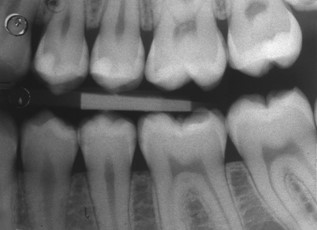
Panoramic bite-wing X-ray – indicated for viewing lateral zones with a single exposure covering all four quadrants.
Indications for bite-wing radiographs are:
– Early detection of approximal dental caries at the contact points of the crown, as well as small cervical caries or caries beneath full-coverage crowns.
– Assessment of bone lesions in marginal periodontopathies, especially at the level of the septum apex and the alveolar ridge.
– Detection of enamel cracks and crown-root fractures resulting from trauma.
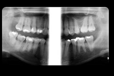
Full-mouth series (FMX) X-ray – includes 18 X-rays using periapical and bite-wing techniques, offering detailed images for all groups of teeth.

Maxillary sinus X-ray – our equipment includes a special program dedicated to maxillary sinuses, allowing us to perform this specialized type of radiologic imaging.
Applicable in fields such as ENT, maxillofacial surgery, and more.
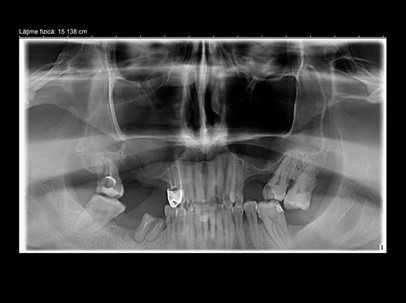
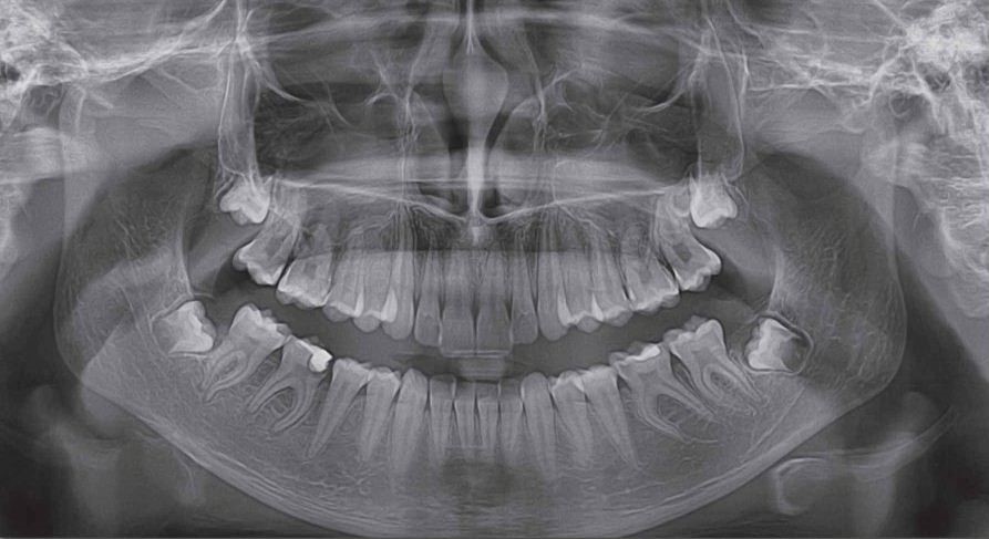
Digital panoramic X-ray (OPT/OPG) – provides a full overview of the dentition and maxillofacial structures; typically performed before starting dental treatment.
Every dentist should recommend a panoramic X-ray at the patient’s first visit before initiating any treatment. Based on the panoramic image, the dentist and patient can discuss the treatment plan — the necessary steps for resolving all existing dental issues.
This type of X-ray captures both dental arches from joint to joint in a single scan, offering a comprehensive panoramic image with low radiation dose (30 µSv) and short scan time (20 seconds).
This investigation is recommended in all dental specialties:
– Orthodontics – for identifying dento-maxillary developmental anomalies, asymmetries, occlusal disturbances, missing teeth, impacted or supernumerary teeth
– Pediatric dentistry – for evaluating tooth eruption and detecting eruption disturbances
– Periodontology – to analyze bone support and tooth implantation, assess the degree and type of alveolar bone resorption
– Implantology – for pre-treatment assessment, identifying all odontogenic or maxillary lesions and anatomical structures that may influence implant placement and osseointegration; also for monitoring implant integration over time, especially in cases with multiple implants across quadrants
– Maxillofacial surgery – for detecting tumors, mandibular and maxillary fractures
– Prosthodontics – in complex oral rehabilitation cases requiring a complete assessment of oral health status
2. 3D COMPUTED TOMOGRAPHY (CBCT)
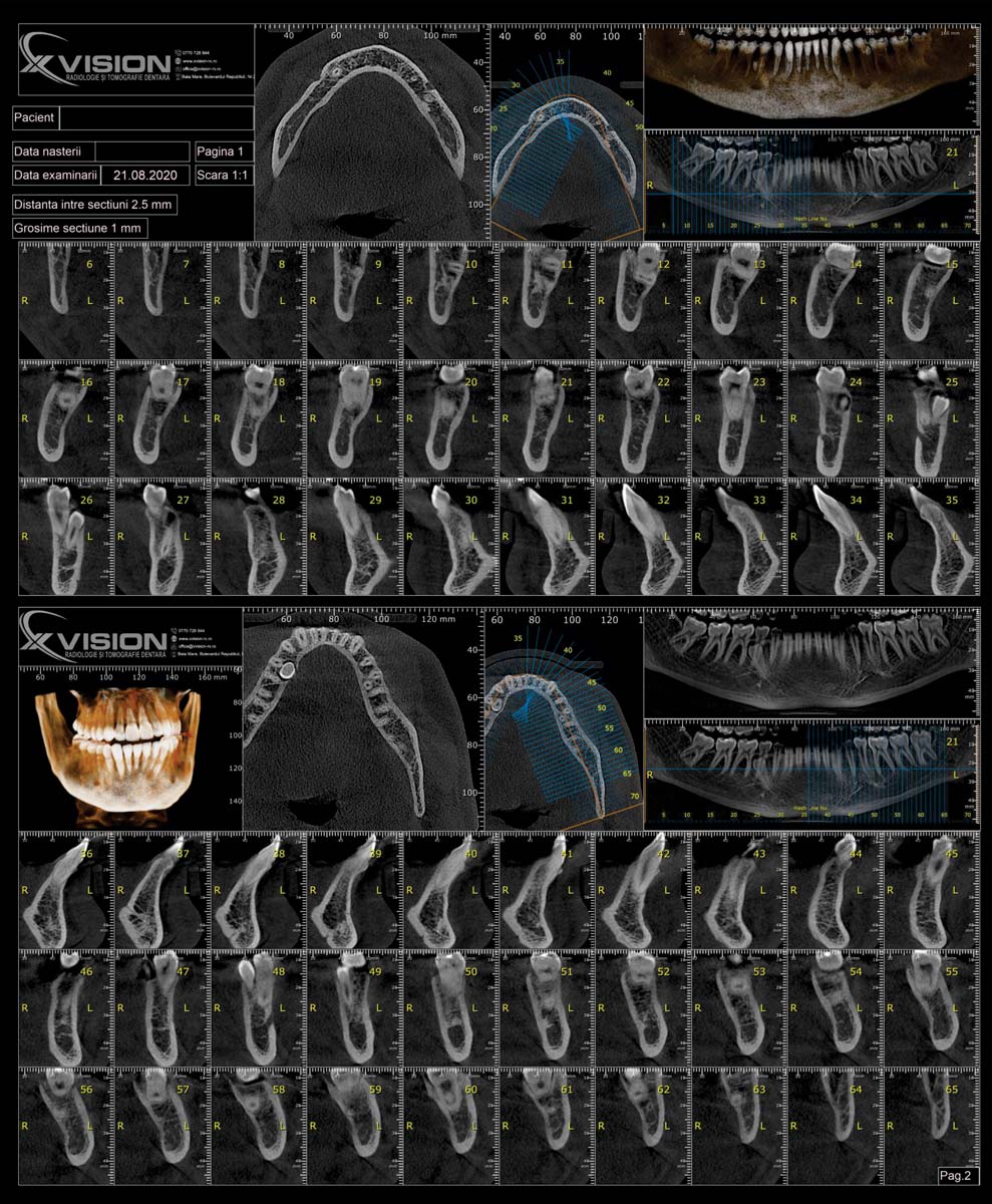
CBCT (Cone Beam Computed Tomography) incorporates cutting-edge imaging technology that allows for three-dimensional visualization of scanned areas.
The CBCT machine looks just like a panoramic X-ray unit, making CT scans much more comfortable compared to traditional fan beam volumetric CTs, which require the patient to lie down or stand still inside the device.
CBCT uses a single, small, cone-shaped X-ray beam. The area of interest is precisely selected by the operator based on the dentist’s recommendation (scanning a group of teeth, a hemi-arch or full arch, or the entire dentition). The scanner requires only a few seconds and a single rotation around the patient’s head to obtain a high-quality 3D image. The use of a cone beam, the limited area of interest, and the short radiation exposure time make the radiation dose 10 to 20 times lower than that of traditional fan beam volumetric CTs.
CBCT and Dental Implants
CBCT analysis allows the implantologist to create a predictable treatment plan. Using dedicated software, future implants can be virtually positioned, minimizing the risk of unforeseen complications during surgery.
CBCT provides critical information about the quantity and quality of existing bone, implant positioning relative to adjacent teeth and cortical bone, nearby anatomical structures and their variations, the presence of pathological formations in the jawbones (granulomas, cysts, tumors).
CBCT has become the standard for clinical decision-making in most dental procedures, such as:
– Deciding whether to extract or treat a tooth
– Precise evaluation of implant placement sites
– Creating surgical implant guides
– TMJ (Temporomandibular Joint) evaluation
– Dynamic assessment of impacted teeth (in rotation)
– Endodontics – visualizing the number, shape, and length of root canals, the quality of endodontic fillings, and identifying apical or periradicular processes
– Periodontology – assessing bone resorption, interradicular lesions, infection pockets
– Evaluating maxillary sinuses and the posterior superior alveolar artery
– Assessing dental and bone trauma
– Mapping the vascular anatomy of the mandibular symphysis and evaluating the lingual mandibular foramen
– Monitoring pathological processes dynamically (in rotation), identifying sclerosis, osteitis, cysts, impacted teeth, and their relationships with neighboring structures (nasal fossae, maxillary sinuses, mandibular canal, etc.)

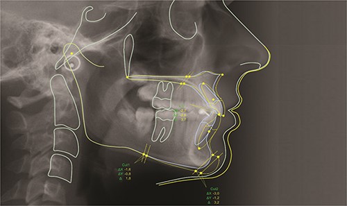
3. DIGITAL CEPHALOMETRY
Lateral cephalometric radiograph – the most accurate investigation for diagnosing dento-maxillary anomalies.
It provides information about the growth direction and development of bone structures.

Postero-anterior cephalometric radiograph – offers insights into the vertical and transverse development of the facial structure.
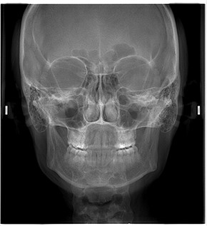
SAF – Fronto-maxillary sinuses
This radiological scan complements the information provided by the maxillary sinus radiograph, especially when the patient’s condition is complex.
The scan is performed using the cephalometric arm of our radiology unit, with special patient positioning.

Digital Cephalometric Analysis with Software
This is the technique of measuring cranial dimensions, commonly used in anthropometric analysis.
With specialized software, any type of cephalometric analysis can be performed using minimal elements such as points or planes.
Photographs, radiographs, analyses, overlays, simulations, animations, and all other document types are organized transparently based on the stages of treatment. They can be used to develop personalized treatment plans for patients.