

CBCT Technology
Cone Beam Computed Tomography
CBCT is a recently developed technology that captures images using a robotic arm that rotates around the patient’s head. This arm holds both the X-ray source and a detector.
The cone-shaped X-rays are directed through the center of the patient’s head to the detector on the opposite side. Both the X-ray source and the detector perform a full or partial rotation, during which between 150 and 600 sequential, flat images are captured. These images are then digitally reconstructed into a complete 3D image.
Complex software is then used by doctors to manipulate the image in any way necessary, allowing them to view the interior of any structure captured by the scan.
The images obtained are useful in dental treatments, surgical interventions, and in diagnosing sinus and auditory canal conditions.
Unlike 2D digital dental X-rays, cone beam CT provides complete three-dimensional images without distortion or overlapping and allows for much more precise measurements.
While X-rays can only be enlarged to analyze details, dental CT scans can be digitally manipulated in specially designed software to be rotated and viewed from any angle.
The appearance of cone beam dental CT, dedicated specifically to dental use, marked a revolution in dentistry, moving from 2D to 3D imaging.
Virtual tissue sections can also be created to determine various characteristics.
Thanks to the cephalometric arm of the KaVo OP 3D device at X Vision, orthodontists can assess lateral X-rays, providing all diagnostic details necessary for orthodontic or orthognathic surgery treatment.
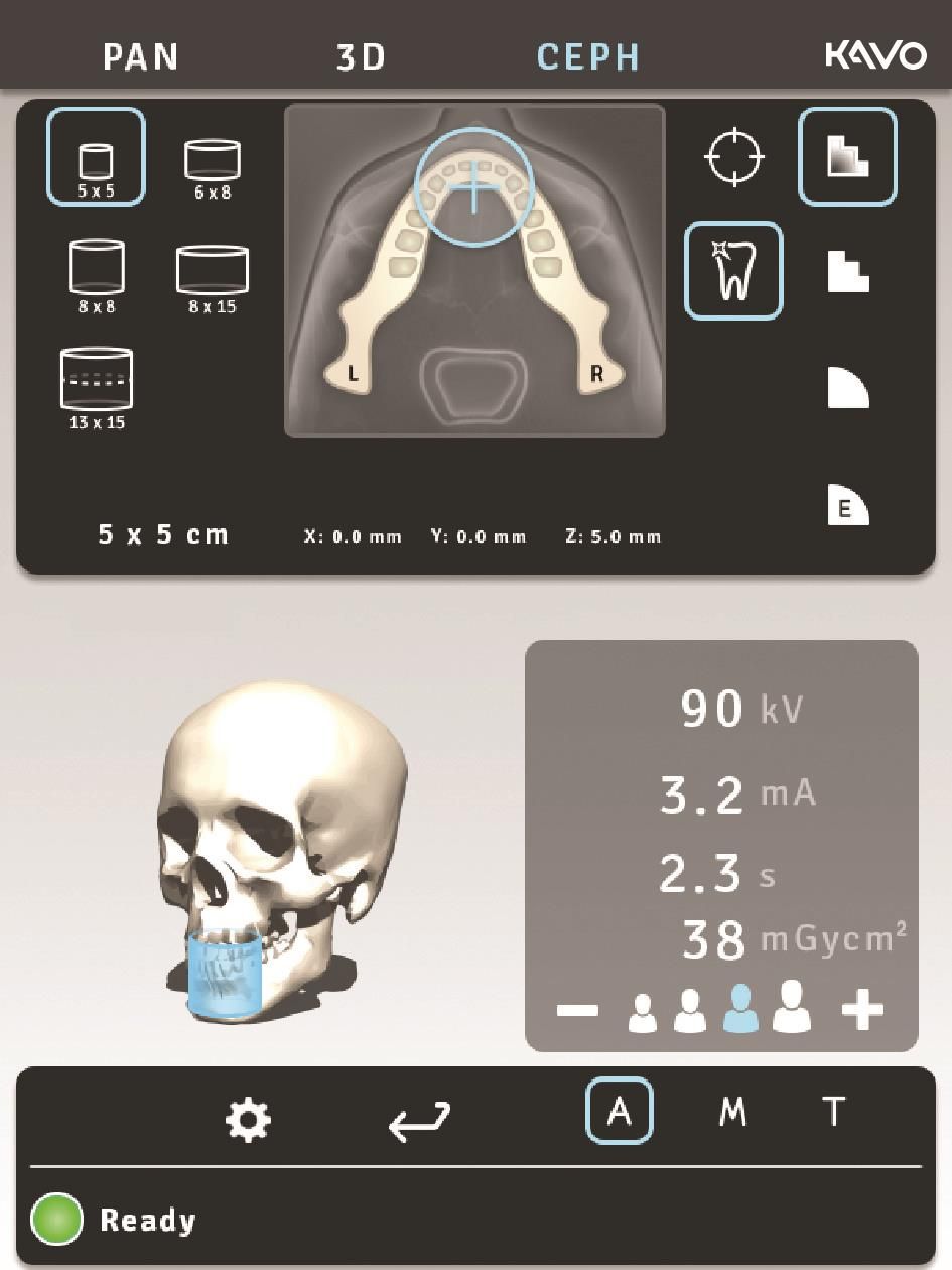
CBCT — short for Cone Beam Computed Tomography—is used in dental fields requiring clear images of bone structures, such as oral and maxillofacial surgery, endodontics, orthodontics, and especially implantology. One of the most important features of this technology is the simulation of dental implants.
Dental CT is used for more complex cases:
- diagnosing more difficult cases
- In implantology treatments:
- Observing bone density in the future implant site and determining whether implantation is possible or if additional procedures such as bone grafting are needed
- simulating implant insertion
- verifying the osseointegration of implants after treatment
- before any maxillofacial surgery
- in complex endodontic treatments, to volumetrically (3D) and accurately visualize the number of canals, their shape and direction, and any root fractures or cracks that may not be visible on a 2D X-ray
- in orthodontics, for 3D visualization of anatomical structures, bone symmetry, and airways
- studying the sinuses
What are the advantages?
- Helps establish an accurate and safe diagnosis – much better visibility and clarity
- Provides information about facial symmetry
- Identifies tumors and cysts in the maxillary area
- Clear imaging of sinuses and polyps
- Simulates the treatment procedure
- Diagnosis of periodontitis
- Helps determine the appropriate type of dental implant and simulates its insertion
- Ensures the implant fits perfectly and reduces intervention time
- Analyzes bone density and simulates bone reconstruction
- Much lower radiation dose compared to conventional CT scans
When is CBCT recommended?
- Confirming the presence of tumors
- Preparing for implant placement
- Before bone grafting surgeries – confirms their necessity and simulates the procedure
- For an accurate analysis of the overall condition of your dentition
- Observing the development of mixed dentition in children
- Before orthodontic treatments, to examine dental arches and jawbones in detail
- During orthodontic and periodontal treatments, to accurately monitor results and progress
- For planning extractions and any type of surgical intervention
- For the correct execution of endodontic treatments (cavities and root canal therapy)
- Detecting fractures
